Difference between revisions of "Projects"
| Line 28: | Line 28: | ||
------- | ------- | ||
| + | ---- /!\ '''Edit conflict - other version:''' ---- | ||
| + | |||
| + | ---- /!\ '''Edit conflict - your version:''' ---- | ||
| + | === Augmented Reality Navigation System for Laparoscopic Prostatectomy === | ||
| + | [[Media:Projects$LaparoscopicProstatectomy.png|[[Media:Projects$LaparoscopicProstatectomy.png|Screenshot|of Augmented Reality visualization during laparoscopic prostatectomy|width="200",align="right"]]]] | ||
| + | |||
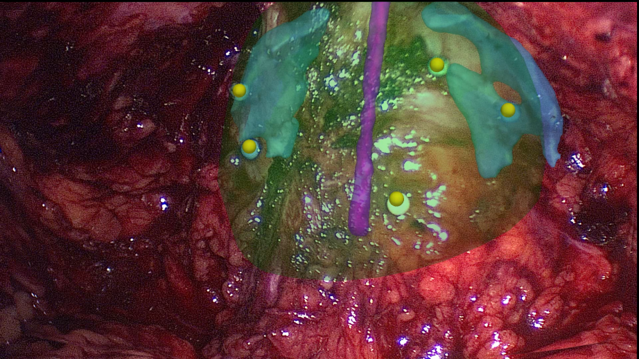
| + | '''Background:''' In laparoscopic radical prostatectomy (LRP) an accurate preparation near the organ may spare the patient’s pelvic innervation and consequently preserve urinal continence and sexual potency. To support the dissection of the prostate an Augmented Reality (AR) Navigation System which conveys virtual organ models generated from transrectal ultrasonography (TRUS) onto the real endoscopic video during radical prostatectomy was created using MITK. The system utilizes custom-developed needles with colored heads which are inserted into the prostate as soon as the organ surface is uncovered. A 3D TRUS scan is acquired right after the placement of the needles and processed using MITK segmentation framework to delineate the important structures (nerve bundles, urethra, etc.). Furthermore, the heads of the needles are located in the 3D ultrasound using the interactive point setting tools in MITK. Thereafter, the system traces the navigation aids in real-time and correctly superimposes the TRUS-based 3D information on an additional AR monitor placed next to the normal endoscopic screen. As a result, the surgeon is provided with valuable information of hidden structures and enables a safe and efficient resection of the prostate. [http://www.dkfz.de/en/mbi/projects/prostata.html Read more ...] | ||
| + | <span style="font-size: smaller">Related publication: Simpfendörfer T, Baumhauer M, Müller M, Gutt CN, Meinzer HP, Rassweiler JJ, Guven S, Teber D. Augmented reality visualization during laparoscopic radical prostatectomy. Journal of Endourology, Volume 25, 2011</span> | ||
| + | |||
| + | ------- | ||
| + | |||
| + | ---- /!\ '''End of edit conflict''' ---- | ||
=== MITK Diffusion Imaging === | === MITK Diffusion Imaging === | ||
'''Background:''' The MITK Diffusion application offers a selection of image analysis algorithms for the processing of diffusion-weighted MR images. It encompasses the research of the Neuroimaging Group in the Division Medical and Biological Informatics at the German Cancer Research Center (dkfz). See [[DiffusionImaging]] for more info. | '''Background:''' The MITK Diffusion application offers a selection of image analysis algorithms for the processing of diffusion-weighted MR images. It encompasses the research of the Neuroimaging Group in the Division Medical and Biological Informatics at the German Cancer Research Center (dkfz). See [[DiffusionImaging]] for more info. | ||
--------- | --------- | ||
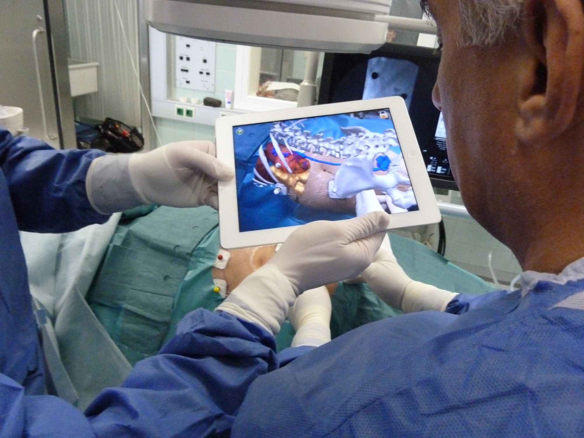
| + | === Using MITK for Augmented Reality Navigation on Mobile Devices === | ||
| + | [[Media:Projects$TabletNavigation.jpg|[[Media:Projects$TabletNavigation.jpg|Screenshot|of tablet navigation during percutaneous nephrolithotomy|width="200",align="right"]]]] | ||
| + | |||
| + | '''Background:''' On basis of MITK an Augmented Reality (AR) navigation system was developed which utilizes a conventional tablet PC to superimpose a transparent, virtual model of a patient gained from CT images onto the video stream recorded by the tablet's camera. As a result, a virtual insight into the patient is generated. | ||
| + | The real-time registration of the 2D video images with the 3D CT content is done utilizing radio-dense, colored markers, easily detectable in both imaging modalities. After affixing the markers on the patient's skin, a CT scan is performed. Prior to the intervention, the CT image is segmented using the MITK interactive segmentation tools to create a three-dimensional, virtual model of the patient. The tablet is equipped with MITK MES (MITK for mobile devices) which was recently developed. The tablet captures images of the surgical site and sends them via Wi-Fi to a tracking server which runs the MITK Workbench. The Workbench in turn runs the registration routines and creates and sends the rendered AR images back to the tablet where they are displayed. The system is currently evaluated in percutaneous nephrolithotomy which is a procedure to remove kidney stones. | ||
| + | |||
| + | <span style="font-size: smaller">Related publication: Simpfendörfer T, Baumhauer M, Müller M, Gutt CN, Meinzer HP, Rassweiler JJ, Guven S, Teber D. Augmented reality visualization during laparoscopic radical prostatectomy. Journal of Endourology, Volume 25, 2011</span> | ||
| + | ------- | ||
=== Statistical tracking of tree-like tubular structures === | === Statistical tracking of tree-like tubular structures === | ||
| Line 41: | Line 61: | ||
------- | ------- | ||
| + | ---- /!\ '''Edit conflict - other version:''' ---- | ||
| + | |||
| + | ---- /!\ '''Edit conflict - your version:''' ---- | ||
| + | === Navigated Bronchoscopy === | ||
| + | |||
| + | [[Media:Projects$NaviBroncho.png|[[Media:Projects$NaviBroncho.png|Screenshot|of TBNA |width="250",align="right"]]]] | ||
| + | |||
| + | '''Background:''' Bronchoscopic interventions, such as transbronchial needle aspiration (TBNA), are commonly performed procedures to diagnose and stage lung cancer. However, due to the complex structure of the lung, one of the main challenges is to find the exact position to perform a biopsy and to actually hit the biopsy target. | ||
| + | To overcome this problem an image-guided, electromagnetic navigation system for transbronchial interventions was developed. Utilizing MTIK and MITK-IGT, the system provides real time positioning information for the bronchoscope and a transbronchial biopsy instrument with only one preoperatively acquired computed tomography image. A twofold respiratory motion compensation method based on a particle filtering approach further allows for guidance through the entire respiratory cycle. [http://www.dkfz.de/en/mbi/projects/bronchoskopie.html Read more ...] | ||
| + | |||
| + | <span style="font-size: smaller">Related publication: Gergel I, Gaa J, Müller M, Meinzer HP, Wegner I. A novel fully automatic system for the evaluation of electromagnetic tracker. In: Medical Imaging 2012: Image-Guided Procedures, Robotic Interventions, and Modeling / David R. Holmes III, Kenneth H. Wong, SPIE Bellingham, Washington, 2012. </span> | ||
| + | |||
| + | ------- | ||
| + | |||
| + | ---- /!\ '''End of edit conflict''' ---- | ||
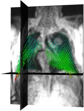
=== Dynamic Analysis of Respiratory Movement === | === Dynamic Analysis of Respiratory Movement === | ||
| Line 54: | Line 89: | ||
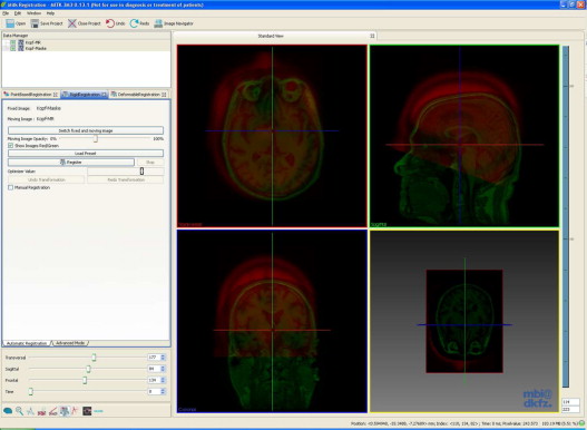
'''Background:''' Although non-rigid registration methods are available or under development for many specific problems in medicine, rigid and affine registration is an important task that is often performed for pre-aligning images before using non-rigid registration. We present a free and open-source application for rigid and affine image registration, which is designed both for developers and for end-users. The application is based on the MITK and allows for inter-modality and intra-modality rigid 2D-2D and 3D-3D registration of medical images such as CT, MRI, or ultrasound. The framework as well as the application can be easily extended by adding new transforms, metrics and optimizers. Additionally, the framework provides the possibility to use image masks to restrict the evaluation of metric values by the optimizer on certain areas of the images. | '''Background:''' Although non-rigid registration methods are available or under development for many specific problems in medicine, rigid and affine registration is an important task that is often performed for pre-aligning images before using non-rigid registration. We present a free and open-source application for rigid and affine image registration, which is designed both for developers and for end-users. The application is based on the MITK and allows for inter-modality and intra-modality rigid 2D-2D and 3D-3D registration of medical images such as CT, MRI, or ultrasound. The framework as well as the application can be easily extended by adding new transforms, metrics and optimizers. Additionally, the framework provides the possibility to use image masks to restrict the evaluation of metric values by the optimizer on certain areas of the images. | ||
| − | <span style="font-size: smaller"> | + | <span style="font-size: smaller">Related publication: Stein D, Fritzsche KH, Nolden M, Meinzer HP, Wolf I. The extensible open-source rigid and affine image registration module of the Medical Imaging Interaction Toolkit (MITK). Comput Methods Programs Biomed, 2010 Oct.</span> |
------- | ------- | ||
| Line 70: | Line 105: | ||
'''Background:''' Navigation systems are promising tools for improving efficacy and safety in surgical endoscopy and other minimally invasive techniques. The aim of this study is to investigate electromagnetic tracking (EMT) for navigated renal access in a porcine model. For our proof-of-principle study we modified a recently established porcine ex vivo model. Via a ureteral catheter which was placed into the desired puncture site, a small sensor was introduced and located by electro magnetic tracking (EMT). Then, a tracked needle was guided into the collecting system in a ‘‘rendezvous’’ approach. A total of 90 renal tracts were obtained in six kidneys using EMT, with a maximum of three punctures allowed per intervention. For each puncture, number of attempts to success, final distance to probe, puncture time, and localization were assessed. We compared absolute and relative frequencies using the chi-square test and applied the Mann–Whitney U-test for continuous variables. No major problems were encountered performing the experiment. Access to the collecting system was successfully obtained after a single puncture in 91% (82/90) and within a second attempt in the remaining 9% (8/90). Thus, a 100% success rate was reached after a maximum of two punctures. With respect to other established techniques, the use of EMT seems to decrease the number of attempts and procedural time remarkably. This might contribute to greater safety and efficacy when applied clinically. | '''Background:''' Navigation systems are promising tools for improving efficacy and safety in surgical endoscopy and other minimally invasive techniques. The aim of this study is to investigate electromagnetic tracking (EMT) for navigated renal access in a porcine model. For our proof-of-principle study we modified a recently established porcine ex vivo model. Via a ureteral catheter which was placed into the desired puncture site, a small sensor was introduced and located by electro magnetic tracking (EMT). Then, a tracked needle was guided into the collecting system in a ‘‘rendezvous’’ approach. A total of 90 renal tracts were obtained in six kidneys using EMT, with a maximum of three punctures allowed per intervention. For each puncture, number of attempts to success, final distance to probe, puncture time, and localization were assessed. We compared absolute and relative frequencies using the chi-square test and applied the Mann–Whitney U-test for continuous variables. No major problems were encountered performing the experiment. Access to the collecting system was successfully obtained after a single puncture in 91% (82/90) and within a second attempt in the remaining 9% (8/90). Thus, a 100% success rate was reached after a maximum of two punctures. With respect to other established techniques, the use of EMT seems to decrease the number of attempts and procedural time remarkably. This might contribute to greater safety and efficacy when applied clinically. | ||
| − | <span style="font-size: smaller"> | + | <span style="font-size: smaller">Related publication: Huber J, Wegner I, Meinzer HP, Hallscheidt P, Hadaschik B, Pahernik S, Hohenfellner M. Navigated renal access using electromagnetic tracking: an initial experience. In Surgical Endoscopy, 2010 Sept.</span> |
Revision as of 15:47, 17 January 2013
Projects using MITK
- The Computer Graphics & Visualization Lab at the Korea Advanced Institute of Science and Technology uses MITK in several projects
- The Centre for Medical Image Computing at the University College London has created NiftyView, a MITK-based working environment for clinical collaborators and research scientists Read more...
MBI-based projects
All projects of the Department of Medical and Biological Informatics at the German Cancer Research Center use MITK for their software development. An overview can be found there.
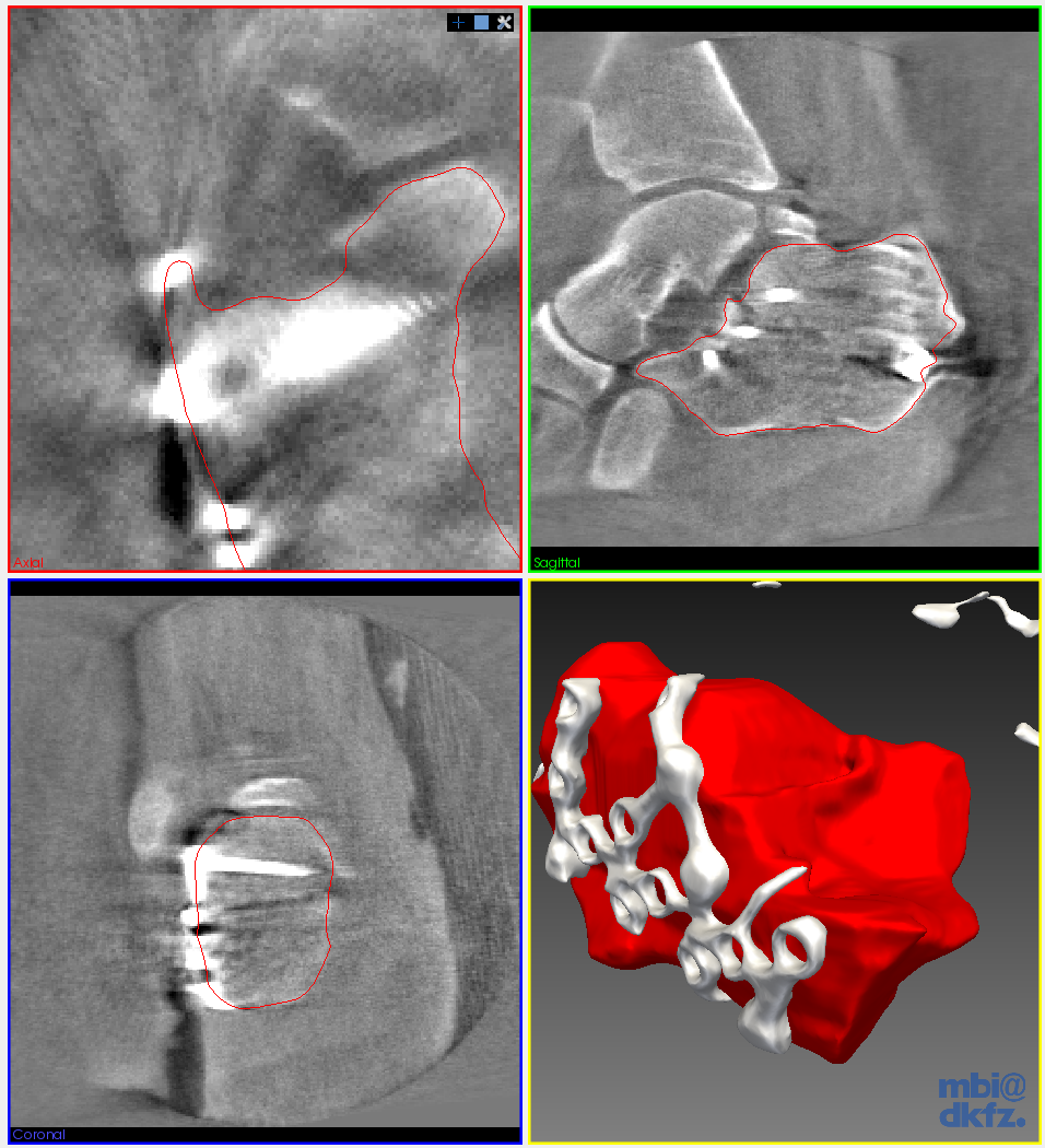
Automated landmark detection and segmentation for intra-operative C-Arm CT applications
[[Media:Projects$HeelBones.png|Screenshot|of 3D c-arms image of heel bone|width="80",align="right"]]
Background: With the development of mobile 3D C-arms, complex bone structures can be visualized during surgery. We design algorithms that facilitate the usability of a mobile 3D imaging system and provide an automatic assessment of a surgical procedure. Thus, working with the imaging system will be faster and allows surgeons to operate with a higher accuracy.
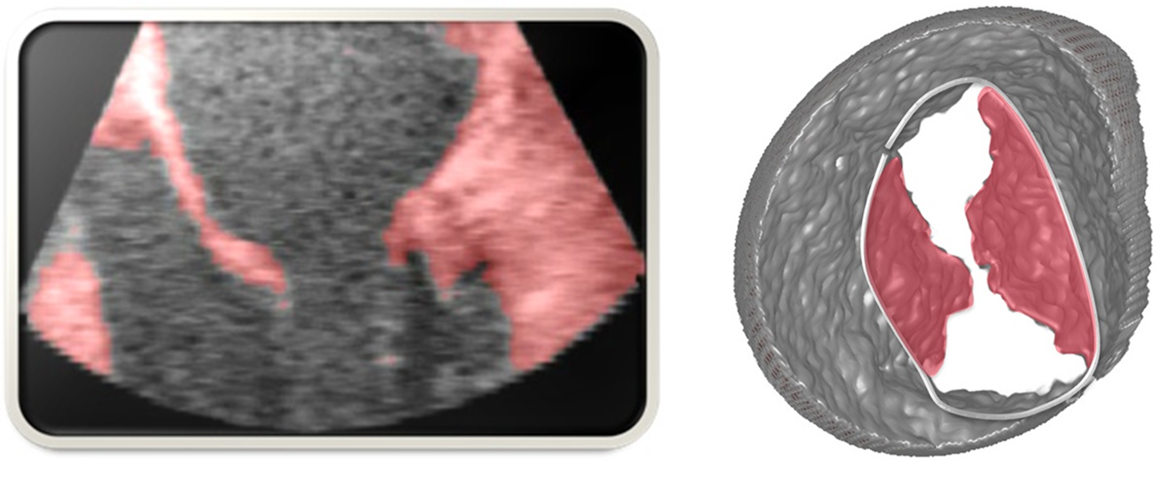
Mitral Valve Modeling based on 4D TEE Data
[[Media:Projects$MitralValveModeling.png|Screenshot|of mitral valve modeling project|width="250",align="right"]]
Background: In mitral valve surgery, the 4D transesophageal echocardiogram has become a standard imaging procedure. Yet, the enormous amount of data produced by this method is hardly exploited because of a lack of respective algorithms. In this project, 4D TEE sequences are used to generate a mathematical model of the mitral valve. This model can be used to perform precise measurements to quantify irregularities, to determine the success after a surgical operation or to compare different patients and treatments. Read more ...
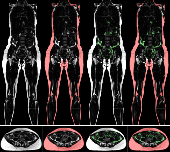
Quantification of Whole-Body Adipose Tissue
[[Media:Projects$AdiposeTissueAnalysis.jpg|Screenshot|of adipose tissue analyse|width="250",align="right"]]
Background: Investigations of prospective studies on the role of overweight and obesity are based mostly on simple metrics such as Body Mass Index (BMI) or waist circumference. The distribution of adipose tissue, which cannot be determined by these simple metrics, is correlated to metabolic syndromes of obesity. For a while, whole-body magnetic resonance imaging (MRI) has been used for a precise assessment of adipose tissue. Compared to the simple metrics, MRI provides an exact amount and the distribution of fat tissue. However, the major advantage of this data is the possibility to quantify the amount of different types of adipose tissue. Three kinds of fat can be distinguished: total, subcutaneous, and visceral adipose tissue. Read more ...
/!\ Edit conflict - other version: ----
/!\ Edit conflict - your version: ----
[[Media:Projects$LaparoscopicProstatectomy.png|Screenshot|of Augmented Reality visualization during laparoscopic prostatectomy|width="200",align="right"]]
Background: In laparoscopic radical prostatectomy (LRP) an accurate preparation near the organ may spare the patient’s pelvic innervation and consequently preserve urinal continence and sexual potency. To support the dissection of the prostate an Augmented Reality (AR) Navigation System which conveys virtual organ models generated from transrectal ultrasonography (TRUS) onto the real endoscopic video during radical prostatectomy was created using MITK. The system utilizes custom-developed needles with colored heads which are inserted into the prostate as soon as the organ surface is uncovered. A 3D TRUS scan is acquired right after the placement of the needles and processed using MITK segmentation framework to delineate the important structures (nerve bundles, urethra, etc.). Furthermore, the heads of the needles are located in the 3D ultrasound using the interactive point setting tools in MITK. Thereafter, the system traces the navigation aids in real-time and correctly superimposes the TRUS-based 3D information on an additional AR monitor placed next to the normal endoscopic screen. As a result, the surgeon is provided with valuable information of hidden structures and enables a safe and efficient resection of the prostate. Read more ... Related publication: Simpfendörfer T, Baumhauer M, Müller M, Gutt CN, Meinzer HP, Rassweiler JJ, Guven S, Teber D. Augmented reality visualization during laparoscopic radical prostatectomy. Journal of Endourology, Volume 25, 2011
/!\ End of edit conflict ----
MITK Diffusion Imaging
Background: The MITK Diffusion application offers a selection of image analysis algorithms for the processing of diffusion-weighted MR images. It encompasses the research of the Neuroimaging Group in the Division Medical and Biological Informatics at the German Cancer Research Center (dkfz). See DiffusionImaging for more info.
[[Media:Projects$TabletNavigation.jpg|Screenshot|of tablet navigation during percutaneous nephrolithotomy|width="200",align="right"]]
Background: On basis of MITK an Augmented Reality (AR) navigation system was developed which utilizes a conventional tablet PC to superimpose a transparent, virtual model of a patient gained from CT images onto the video stream recorded by the tablet's camera. As a result, a virtual insight into the patient is generated. The real-time registration of the 2D video images with the 3D CT content is done utilizing radio-dense, colored markers, easily detectable in both imaging modalities. After affixing the markers on the patient's skin, a CT scan is performed. Prior to the intervention, the CT image is segmented using the MITK interactive segmentation tools to create a three-dimensional, virtual model of the patient. The tablet is equipped with MITK MES (MITK for mobile devices) which was recently developed. The tablet captures images of the surgical site and sends them via Wi-Fi to a tracking server which runs the MITK Workbench. The Workbench in turn runs the registration routines and creates and sends the rendered AR images back to the tablet where they are displayed. The system is currently evaluated in percutaneous nephrolithotomy which is a procedure to remove kidney stones.
Related publication: Simpfendörfer T, Baumhauer M, Müller M, Gutt CN, Meinzer HP, Rassweiler JJ, Guven S, Teber D. Augmented reality visualization during laparoscopic radical prostatectomy. Journal of Endourology, Volume 25, 2011
Statistical tracking of tree-like tubular structures
[[Media:Projects$TrackingTubularStructure.png|Screenshot|of vessel segmentation|width="250",align="right"]]
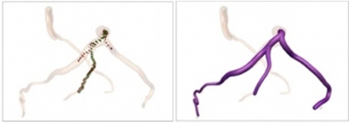
Background: The segmentation of tubular structures from 3D medical image data is the essential basis for many computer-assisted applications such as operation planning and the development of an individualized ventilation strategy. Our project has as its objective the development of a robust segmentation procedure to achieve precise results with few user interactions in a clinically feasible amount of time. Read more ...
/!\ Edit conflict - other version: ----
/!\ Edit conflict - your version: ----
[[Media:Projects$NaviBroncho.png|Screenshot|of TBNA |width="250",align="right"]]
Background: Bronchoscopic interventions, such as transbronchial needle aspiration (TBNA), are commonly performed procedures to diagnose and stage lung cancer. However, due to the complex structure of the lung, one of the main challenges is to find the exact position to perform a biopsy and to actually hit the biopsy target. To overcome this problem an image-guided, electromagnetic navigation system for transbronchial interventions was developed. Utilizing MTIK and MITK-IGT, the system provides real time positioning information for the bronchoscope and a transbronchial biopsy instrument with only one preoperatively acquired computed tomography image. A twofold respiratory motion compensation method based on a particle filtering approach further allows for guidance through the entire respiratory cycle. Read more ...
Related publication: Gergel I, Gaa J, Müller M, Meinzer HP, Wegner I. A novel fully automatic system for the evaluation of electromagnetic tracker. In: Medical Imaging 2012: Image-Guided Procedures, Robotic Interventions, and Modeling / David R. Holmes III, Kenneth H. Wong, SPIE Bellingham, Washington, 2012.
/!\ End of edit conflict ----
Dynamic Analysis of Respiratory Movement
Background: In the course of numerous lung diseases, and following therapy, a reduction in respiratory motion and thus restricted pulmonary function is observed. New imaging techniques have recently provided non-invasive methods for depicting these changes. The aim of the project is to establish the technical basis for analyzing respiratory motion using 3D+t magnetic resonance imaging or computed tomography. To this end, one of the approaches currently being followed is to define vector fields that approximately reflect the motion of lung parenchyma during the respiratory cycle. The analysis of the vector fields permits conclusions about the movement, within a local area, and deformation of tissue. When coded onto a color map, this information may be used, for instance, to track how successful a therapy is over time. Read more ...
The extensible open-source rigid and affine image registration module of the Medical Imaging Interaction Toolkit (MITK)
Media:Projects$stein-registration.jpeg|Screenshot|of registration module|width="250",align="right"
Background: Although non-rigid registration methods are available or under development for many specific problems in medicine, rigid and affine registration is an important task that is often performed for pre-aligning images before using non-rigid registration. We present a free and open-source application for rigid and affine image registration, which is designed both for developers and for end-users. The application is based on the MITK and allows for inter-modality and intra-modality rigid 2D-2D and 3D-3D registration of medical images such as CT, MRI, or ultrasound. The framework as well as the application can be easily extended by adding new transforms, metrics and optimizers. Additionally, the framework provides the possibility to use image masks to restrict the evaluation of metric values by the optimizer on certain areas of the images.
Related publication: Stein D, Fritzsche KH, Nolden M, Meinzer HP, Wolf I. The extensible open-source rigid and affine image registration module of the Medical Imaging Interaction Toolkit (MITK). Comput Methods Programs Biomed, 2010 Oct.
Media:Projects$ablation1.jpg|Ablation|planning|width="250",align="right"
Background: Minimally invasive procedures for cancer diagnosis and therapy are increasingly replacing open procedures in the clinical routine because of their protective character. Therapeutic thermal procedures such as laser-induced thermotherapy (LITT), radiofrequency ablation (RFA) and cryotherapy are, for example, increasingly being used in the minimally invasive treatment of hepatic tumors. The common underlying principle of these procedures is tissue destruction by means of local temperature changes. The success of the intervention crucially depends on the precision of the instrument insertion and thus the experience of the physician. After computer-assisted navigation on rigid structures has become an established practice in the clinical routine, the use of computer support in soft tissues has become limited to non-invasive diagnostics and operation planning. This especially can be attributed to the absent compensation of intra-interventional organ movement. The objective of this project is to develop, implement and evaluate new concepts for computer-assisted biopsies in soft tissues.
[[Media:Projects$ToolPairNavigation.jpeg|Description|width="250",align="right"]]
Background: Navigation systems are promising tools for improving efficacy and safety in surgical endoscopy and other minimally invasive techniques. The aim of this study is to investigate electromagnetic tracking (EMT) for navigated renal access in a porcine model. For our proof-of-principle study we modified a recently established porcine ex vivo model. Via a ureteral catheter which was placed into the desired puncture site, a small sensor was introduced and located by electro magnetic tracking (EMT). Then, a tracked needle was guided into the collecting system in a ‘‘rendezvous’’ approach. A total of 90 renal tracts were obtained in six kidneys using EMT, with a maximum of three punctures allowed per intervention. For each puncture, number of attempts to success, final distance to probe, puncture time, and localization were assessed. We compared absolute and relative frequencies using the chi-square test and applied the Mann–Whitney U-test for continuous variables. No major problems were encountered performing the experiment. Access to the collecting system was successfully obtained after a single puncture in 91% (82/90) and within a second attempt in the remaining 9% (8/90). Thus, a 100% success rate was reached after a maximum of two punctures. With respect to other established techniques, the use of EMT seems to decrease the number of attempts and procedural time remarkably. This might contribute to greater safety and efficacy when applied clinically.
Related publication: Huber J, Wegner I, Meinzer HP, Hallscheidt P, Hadaschik B, Pahernik S, Hohenfellner M. Navigated renal access using electromagnetic tracking: an initial experience. In Surgical Endoscopy, 2010 Sept.