DiffusionImaging
MITK Diffusion
Quicklinks
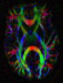
The MITK Diffusion application [1,2] offers a selection of image analysis algorithms for the processing of diffusion-weighted MR images. It encompasses the research of the Division Medical Image Computing at the German Cancer Research Center (DKFZ).
Features & Highlights
- Support for most established image formats, such as DICOM, NIFTI, and NRRD
- Tensor and Q-ball reconstruction
- Intravoxel Incoherent Motion (IVIM) analysis
- Fiber tractography and fiber processing
- Global tractography [3]
- Interactive (similar to [4]) deterministic and probabilistic peak, ODF and tensor streamline tractography
- Machine learning based tractography [5]
- Brain network statistics and visualization (connectomics)
- Fiberfox: simulation of diffusion-weighted MR images [6]
- Command line tools for most functionalities
The MITK Diffusion application is based on the MITK research platform and is completely open-source. The code is embedded into the source code of MITK as a module and can be accessed through the public git repository.
Downloads
If you encounter any bugs, please report them in our bugtracking system or use the MITK-users mailing list. We are grateful for any feedback!
Latest stable installers (2017.07)
Commit hash 2bda849ee86a362583ee3d2beb4baaca038bd8a5
| Windows 7, Windows 10 | MS Windows (64 bit) installer |
| Windows 7, Windows 10 | MS Windows (64 bit) zip archive |
| Ubuntu 16.04 and newer | Ubuntu (64 bit), tar.gz archive |
| Ubuntu 14.04 | Ubuntu (64 bit), tar.gz archive |
Requirements
- For Ubuntu users:
- Install Python 3.X:
sudo apt install python3 python3-pip
- Download Python requirements file: https://phabricator.mitk.org/file/data/r7yikza26ozkodxlypsq/PHID-FILE-thn2yotzr2jein4apf3x/PythonRequirements.txt
- Install Python requirements:
pip3 install -r PythonRequirements.txt
- If your are behind a proxy use
pip3 --proxy <proxy> install -r PythonRequirements.txt
- Install Python 3.X:
- For Windows users:
- MITK Diffusion requires the Microsoft Visual C++ 2017 Redistributable to be installed on the system. The MITK Diffusion installer automatically installs this redistributable for you if not already present on the system, but it needs administrative privileges to do so. So to install the redistributable, run the MITK Diffusion installer as administrator.
- Install Python 3.X: https://www.anaconda.com/download/
- Download Python requirements https://phabricator.mitk.org/file/data/r7yikza26ozkodxlypsq/PHID-FILE-thn2yotzr2jein4apf3x/PythonRequirements.txt
- Install Python requirementsfrom the conda command prompt:
pip install -r PythonRequirements.txt
- If your are behind a proxy use
pip --proxy <proxy> install -r PythonRequirements.txt
- Requirements for all deep-learning based functionalities:
- Affected functionalities:
- Brain extraction
- TractSeg
- Pytorch: https://pytorch.org/
- CUDA: https://developer.nvidia.com/cuda-downloads
- (optional) cuDNN: https://developer.nvidia.com/cudnn
- Affected functionalities:
Known issues
The Fiberfox command line application does not read the b-value but uses a default b-value of 1000 s/mm². This bug does not affect the GUI version of Fiberfox. This bug also has no effect if the first non-zero b-value is 1000 s/mm², which is for example the case in the simulated HCP dataset (10.5281/zenodo.572345). This bug is fixed in the current master of the MITK source code.
Building MITK Diffusion from source
- Install Qt on your system.
- Clone MITK from out git repository using Git version control.
- Configure the MITK Superbuild using CMake.
- Choose the source code directory and an empty binary directory.
- Click "Configure".
- Set the option MITK_BUILD_CONFIGURATION to "DiffusionRelease".
- Click "Generate".
- Build the project
- Linux: Open a console window, navigate to the build folder and type "make -j8" (optionally supply the number threads to be used for a parallel build qith -j).
- Windows (requires visual studio): Open the MITK Superbuild solution file and build all projects.
- The build may take some time and should yield the binaries in "your_build_folder/MITK-build/bin"
More detailed build instructions can be found in the documentation.
References
[1] Fritzsche, Klaus H., Peter F. Neher, Ignaz Reicht, Thomas van Bruggen, Caspar Goch, Marco Reisert, Marco Nolden, et al. “MITK Diffusion Imaging.” Methods of Information in Medicine 51, no. 5 (2012): 441.
[2] Fritzsche, K., and H.-P. Meinzer. “MITK-DI A New Diffusion Imaging Component for MITK.” In Bildverarbeitung Für Die Medizin, n.d.
[3] Neher, P. F., B. Stieltjes, M. Reisert, I. Reicht, H.P. Meinzer, and K. Maier-Hein. “MITK Global Tractography.” In SPIE Medical Imaging: Image Processing, 2012.
[4] Chamberland, M., K. Whittingstall, D. Fortin, D. Mathieu, und M. Descoteaux. „Real-time multi-peak tractography for instantaneous connectivity display“. Front Neuroinform 8 (2014): 59. doi:10.3389/fninf.2014.00059.
[5] Neher, Peter F., Marc-Alexandre Côté, Jean-Christophe Houde, Maxime Descoteaux, and Klaus H. Maier-Hein. “Fiber Tractography Using Machine Learning.” NeuroImage. Accessed July 17, 2017. doi:10.1016/j.neuroimage.2017.07.028.
[6] Neher, Peter F., Frederik B. Laun, Bram Stieltjes, and Klaus H. Maier-Hein. “Fiberfox: Facilitating the Creation of Realistic White Matter Software Phantoms.” Magnetic Resonance in Medicine 72, no. 5 (November 2014): 1460–70. doi:10.1002/mrm.25045.
Contact
If you have questions about the application or if you would like to give us feedback, don't hesitate to contact us using our mailing list or, for questions that are of no interest for the community, directly.
